MICROSCOPIC ENDODONTICS
ON A WORLDWIDE LEVEL AND TOP STANDARDS
The pulp, i.e. the neurovascular bundle, is the heart of our teeth. It is where all vital processes take place – it also alarms us when something is wrong with our tooth. If bacteria get into the tooth and infect the pulp, untreated caries leads to its inflammation. In such situations, root canal treatment is the only way to prevent tooth extraction. The first step in such treatment is removing the inflamed pulp from the root canals and chamber. Next, the root canals are widened using chemical and mechanical techniques, disinfected and filled. This way we are able to remove harmful microorganisms from the root canal system. Nowadays, endodontics is painless: it is performed in anaesthesia, in one or two sessions.

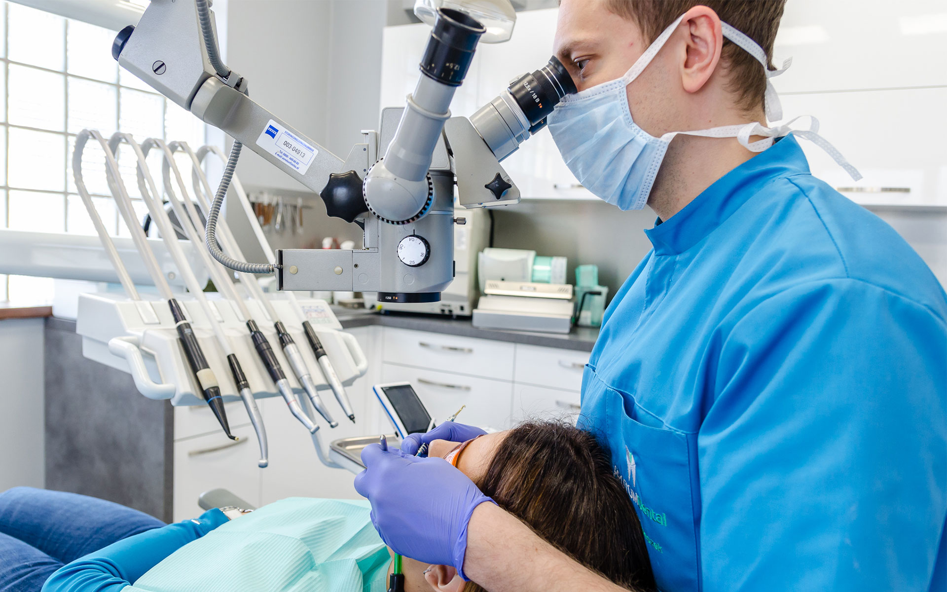
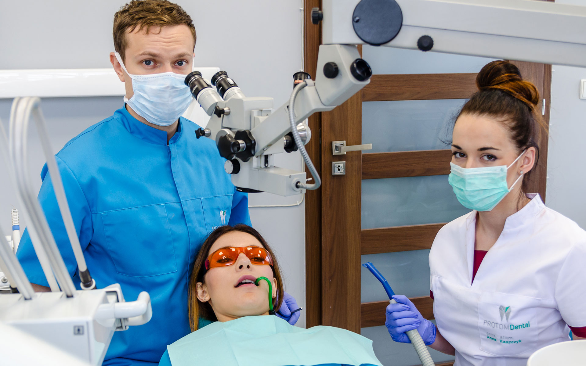
As our services are of the highest quality, we perform endodontics using a microscope. The root canals are pre-treated mechanically and filled with liquid gutta-percha – it is one of the state-of-the-art methods used in dentistry. Precision, safety and effectiveness are the basic advantages of microscopic endodontics. Medical recommendations for this treatment include: curved roots, narrow or fused canals, atypical anatomy of the tooth: any deviation in its anatomy makes it impossible to localise the root canals.
The only way to see the tiniest details is by augmenting the image of the operative site even up to 25 times using a microscope. It determines the course and results of the treatment. The microscope increases the odds of successful treatment and – very often – allows for avoiding tooth extraction. It also supports conservative dentistry when it comes to diagnosing and treating tooth decay: using a microscope allows for removing caries with minimal losses of the healthy tissue.

Before we proceed, we take an x-ray picture of the tooth in question which allows us to specify its anatomy and localise possible periapical lesions. We use modern radiology techniques which ensure safety, clear framing and minimal radiation doses specified individually for each patient. At the end of the treatment we provide our patients with an RVG image. The device is located near the dental chair for the convenience of both our patients and the dentist.
We use the ENDOMETER in root canal treatments. This device measures the length of the root canal with the accuracy of 0.1mm and enhances the odds of successful treatment. Thanks to the endometer there is no need of taking an RVG image in the course of treatment, which is especially important in the case of pregnant patients. At first we isolate the tooth to provide a clean and saliva-free environment, using the so-called dental dam i.e. a rubber sheet placed on the tooth and secured with a clasp.It is more comfortable for the patient than traditional dental lignin rolls. The microscope is placed above the patient’s head allowing for a look into the inside of the tooth which remains open to get the access to the root canals. Success of the treatment depends greatly on the restoration of the tooth. Such therapy weakens the teeth, as they lose even up to a half of their tissue. We replace and reinforce them using dental composites and fiberglass, respectively. Our technical laboratory makes crown-root inlays as well as prosthetic crowns. Our offer also includes the so-called onlays which are made by our technicians and are adjusted to the opposing teeth. Onlays are cemented adhesively during the next visit. Proper treatment and tooth reconstruction are crucial for its durability.


